People
Organizers
Event Coordinator
Speakers
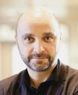
Sylvain Baillet, PhD
Sylvain Baillet is a Professor of Neurology and Neurosurgery, Biomedical Engineering and Computer Science at McGill University. He holds a Tier 1 Canada Research Chair in Neural Dynamics of Brain Systems. In 2019, he was appointed Associate Dean, Research of the Faculty of Medicine. In 2011 he became the founding director of the Magnetoencephalography Brain Imaging core unit (MEG) at the Montreal Neurological Institute and Hospital (The Neuro). Dr. Baillet was Acting Director of The Neuro’s McConnell Brain Imaging Centre from 2013 to 2017. He currently chairs The Neuro’s Open-Science Grassroots Initiatives Committee.
Dr. Baillet’s research focuses on the development of brain imaging methods and quantitative electrophysiology, with an emphasis on MEG, an imaging tool used to map brain activity at the millisecond scale. Primary applications of his research are in systems, cognitive and clinical neuroscience, with an emphasis on the mechanisms of information integration in brain networks, related to complex behavior and symptoms. A co-author of 300 publications, Dr. Baillet is also committed to the promotion of Open Science practices and the development and dissemination of open-source applications for data analytics and data repositories. He co-founded the Brainstorm software project for multimodal brain electrophysiology and imaging, which grew to a community of 25,300 users worldwide. In 2015 he led a Quebec Bioimaging Network strategic initiative to create the Open MEG Archives (OMEGA), the world’s first open-access repository of MEG data.
Contact
Sylvain Baillet, PhD
Professor
sylvain.baillet@mcgill.ca
https://www.mcgill.ca/neuro/sylvain-baillet-phd
Martin Cousineau, MS
Martin Cousineau graduated summa cum laude from the University of Ottawa in mathematics and computer science. He went on to do his Master's in computational neuroimaging at the University of Sherbrooke, working with diffusion MRI at the Sherbrooke Connectivity Imaging Laboratory (SCIL). In 2017, he joined Sylvain Baillet at the McConnel Brain Imaging Centre to help with the Brainstorm development and the new open data initiatives.
Martin Cousineau, MS
Software Developer
Contact
martin.p.cousineau@mcgill.ca
Abhishek Prasad, PhD
Abhishek Prasad received his M.S. in Biomedical Engineering from Louisiana Tech University and Ph.D. in Biomedical Engineering from the New Jersey Institute of Technology and University of Medicine and Dentistry in NJ. He is currently an Assistant Professor in the Department of Biomedical Engineering at the University of Miami. The goal of his Neural Interfaces Lab is to develop brain and spinal cord machine interfaces to restore communication and control in paralyzed individuals. The lab is also involved in neural probe development by understanding and mitigating various material and biological failure mechanisms in neural electrode failure.
Talk Title:
EEG brain computer interface-functional electrical stimulation (BCI-FES) for hand functions in spinal cord injury
Abstract:
In the United States, there are over 33,000 people living with complete tetraplegia due to a traumatic spinal cord injury (SCI). Restoration of hand and arm function is ranked as the highest priority for people living with complete tetraplegia, because it would offer greater independence and improved quality of life. Activation of paralyzed hand muscles by functional electrical stimulation (FES) has been shown to restore control following complete spinal cord injury (SCI). A variety of control signals have been used, but most require head or arm movements, which may interfere with activities of daily living. Alternatively, natural signals from the brain can be used for control. In this study, subjects with chronic (>1 year post-injury) C5/C6 motor-complete SCI control a brain-controlled functional electrical stimulation system for grasp and release. Through these studies, we seek to create a BCI-FES as an assistive system for tetraplegics and study neuroplastic changes that may occur during BCI usage.
Contact
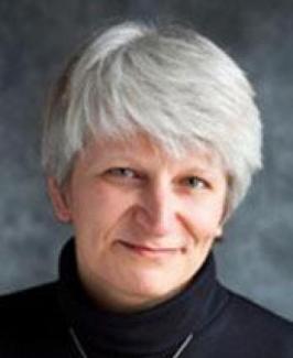
Aina Puce, PhD
Research. In my Social Neuroscience Lab we study the brain basis of non-verbal social cognition – particularly how consciously or unconsciously perceived information helps us assess others’ intentions, goals and mental states. My research integrates functional and structural magnetic resonance imaging (MRI), infrared eye tracking, high-density electroencephalography (EEG), magnetoencephalography (MEG) and electrocorticography (ECoG). By investigating sbrain tructural and functional connectivity patterns as subjects engage in evaluating the social behaviors of others, we aim to develop insights into how our brains processes the continuous daily barrage of dynamic and fleeting incoming social information.
Methods training. Science is important, but must be based on a solid scaffold of valid scientific method. To that end, I have co-written a textbook on MEG-EEG methods [Riitta Hari & Aina Puce (2017) MEG-EEG Primer, New York, Oxford University Press, https://global.oup.com/academic/product/meg-eeg-primer-9780190497774?cc=us&lang=en&]. I am active in an international effort devoted to encouraging best practices in neuroimaging, open science and data analysis/sharing [see Pernet et al., (2018) https://osf.io/a8dhx/].
Talk Title:
Re-using MEEG data and maximizing its value: considerations of statistical power & white matter connectivity
Abstract:
Human neurophysiological data are a rich source of spatio-temporal information. They deepen our understanding of brain-behavior relationships, when combined with other data types [e.g. white matter tractography]. I will present two studies, where MEG data and ECoG data have been re-used to answer specific questions related to experimental design/data analysis or to science itself.
1. MEG and statistical power as a function of number of subjects and trials.
Simulated ‘experiments’ were performed on the MEG resting-state data of 89 subjects from the Human Connectome Project [HCP]. Each subject’s data were divided into two ‘conditions’. In a ‘signal condition’ a dipolar source was injected at a known anatomical location, but not in a ‘noise condition’. First, we detected significant differences at sensor level with classical paired t-tests across subjects, and group-level detectability of simulated effects varied greatly with anatomical origin. Second, we examined which spatial properties of the sources most affected detectability. Not surprisingly, the most detectable effects originate from source positions closest to the sensors with tangential orientation with respect to the sensor array. Also, cross-subject variability in orientation affected group-level effect detectability, aiding detection in regions with small variability and hindering detection where it was large. A considerable covariation of source position, orientation, and cross-subject variability in native brain space was seen, making it difficult to assess effects of these variables independently of one another. Therefore, a second set of similar simulations was performed. This time, spatial properties were controlled independently of individual anatomy, allowing the effects of position, orientation, and cross-subject variability of on detectability to be examined. Results confirmed the strong impact of distance and orientation on source detectability and showed that orientation variability across subjects affects detectability, whereas position variability does not. Study bottom line? [A] Strict unequivocal recommendations as to ideal number of trials and subjects for experiments cannot be realistically provided for MEG studies. [B] Spatial constraints underlying expected sources of activity while planning experiments must be considered at the experimental design stage. This is imperative for network level analyses in cognitive neuroscience studies, where activity in nodes in a network may not be equally well detected. [See: Chaumon M, Puce A, George N. (2020) bioRxiv doi: https://doi.org/10.1101/852202]
2. ECoG, white matter tractography and information flow in face processing pathways.
Intracerebral EEG from 11 epileptic patients were analyzed while they had viewed initially presented neutral faces that turned fearful or happy with, or without, an associated lateral gaze change [amygdala data published as Huijgen et al (2015) doi: 10.1093/scan/nsv048]. N200 field potential peak amplitudes were used for identifying the loci for face processing, and latency served as an indicator of transmission of information within the system. Results indicated that face onset processing begins in inferior occipital cortex and moves, in parallel, anteroventrally to fusiform and inferior temporal cortices. In contrast, the superior temporal sulcus [STS] responded selectively to gaze changes with augmented field potential amplitudes for averted versus direct gaze, and large effect sizes relative to other regions of the network. An overlap analysis of active intracerebral electrodes from the 11 patients with posterior white matter tract endpoints [from 1066 healthy brains, Human Connectome Project] was then performed. Study bottom line? From the combination of ECoG and white matter tract endpoint data it appears that inferior occipital and temporal sulci likely broadcast their information - the former dorsally to intraparietal sulcus, and the latter to both fusiform and superior temporal cortex. These data indicate that: [A] Inferior temporal cortex should be included in future face processing models. [B] Superior temporal cortex is critical for dynamic gaze processing. [See: Babo-Rebelo M, Puce A et al., (2020) submitted*]
Aina Puce, PhD
Associate Professor, Eleanor Cox Riggs Professor
Contact
ainapuce@indiana.edu
http://mypage.iu.edu/~ainapuce/PuceLab/Welcome.html
Jing Xiang, MD, PhD
Dr. Jing Xiang is the Director of Magnetoencephalography (MEG) Research at Cincinnati Children’s Hospital Medical Center. He is an Associate Professor in the Departments of Pediatrics and Neurology at the University of Cincinnati. Dr. Xiang played a key role in building the world’s first pediatric MEG Laboratory at the Hospital for Sick Children, in Toronto, Canada. He also established the world’s first high frequency brain signal database (ClinicalTrials.gov Identifier: NCT00600717). He is a pioneer in clinical applications of high frequency brain signals and has published more than 120 peer-reviewed papers. Dr. Xiang received numerous awards for his outstanding contributions (e.g. Walter Berdon Award).
Talk Title:
High Frequency Brain Signal – Methodology and Application
Abstract:
High frequency brain signals (HFBS) open a new avenue for brain research in normal and abnormal conditions. Invasive recordings have been conventionally used in the study of HFBS. Recent advances in magnetoencephalography (MEG) and scalp electroencephalography (EEG) have shown promising results for noninvasive detection of HFBS. Though invasive recordings can provide convincing data, its applications are limited to accessible brain areas (low spatial sampling) in surgical patients. Modern MEG and EEG can detect HFBS from the entire brain, which open a new window for noninvasive investigation of HFBS for both basic research and clinical application. An important area investigating HFBS is the analyses of functional high gamma signals (> 70 Hz) elicited by tasks or stimuli. High gamma signals have been widely studied in the somatosensory, motor, visual, auditory and language systems. The detection of intrinsic (endogenous) HFBS in the epileptic brain represents another important field of clinical application. Indeed, oscillatory HFBS, such as high frequency oscillations (HFOs, 80-600 Hz), ripples (80-250 Hz), fast ripples (250-600 Hz), and very high frequency oscillations (VHFOs> 600 Hz), have shown promising results in epilepsy surgery. Non-oscillatory HFBS, such as high frequency spikes, have recently been identified, but its clinical usefulness remains largely unknown. This talk will discuss the state of the art methodologies and promising results of clinical applications of HFBS.
Jing Xiang, MD, PhD
Associate Professor
Contact
Jing.Xiang@cchmc.org
https://www.cincinnatichildrens.org/bio/x/jing-xiang


